Human Anatomy Abdomen Female Stomach Drawing - Human Anatomy Abdomen Female Anatomy Drawing Diagram / Jul 26, 2021 · abdominal wall peritoneum stomach spleen liver pancreas small intestine large intestine kidneys and ureters nerves, vessels and lymphatics of the abdomen pelvis and perineum pelvic girdle and floor female pelvis and reproductive organs male pelvis and reproductive organs urinary bladder and urethra perineum nerves, vessels and lymphatics of the.
Human Anatomy Abdomen Female Stomach Drawing - Human Anatomy Abdomen Female Anatomy Drawing Diagram / Jul 26, 2021 · abdominal wall peritoneum stomach spleen liver pancreas small intestine large intestine kidneys and ureters nerves, vessels and lymphatics of the abdomen pelvis and perineum pelvic girdle and floor female pelvis and reproductive organs male pelvis and reproductive organs urinary bladder and urethra perineum nerves, vessels and lymphatics of the.. Gastrointestinal anatomy stomach pyloric sphincter duodenum figs. Somatostatin cells in both the antrum and the fundus. The stomach and duodenum are separated by a short muscular sphincter, the pylorus. It is generally used for making small incisions in skin and muscle. It is usually treated with antibiotics, although may require surgical drainage in complicated cases.
It is generally used for making small incisions in skin and muscle. The stomach and duodenum are separated by a short muscular sphincter, the pylorus. The inside of the left kidney shows the renal pelvis. An inset shows the renal tubules and urine. It is usually treated with antibiotics, although may require surgical drainage in complicated cases.
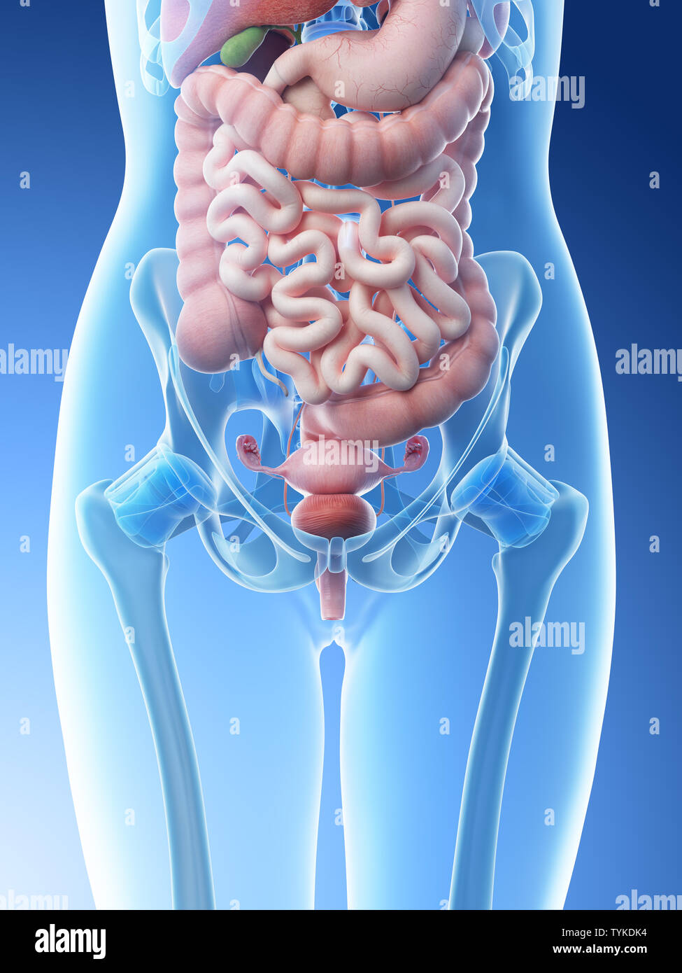
Abdominal Organs High Resolution Stock Photography And Images Alamy from c8.alamy.com Somatostatin cells in both the antrum and the fundus. Jul 26, 2021 · abdominal wall peritoneum stomach spleen liver pancreas small intestine large intestine kidneys and ureters nerves, vessels and lymphatics of the abdomen pelvis and perineum pelvic girdle and floor female pelvis and reproductive organs male pelvis and reproductive organs urinary bladder and urethra perineum nerves, vessels and lymphatics of the. Anatomy of the male urinary system (left panel) and female urinary system (right panel); Sitting the dog on the table or floor, while controlling the head with your hands. An inset shows the renal tubules and urine. Serratus anterior the serratus anterior, as its name suggests, consists of multiple muscle slips that run along the anterolateral chest wall (see figure 1 for surface anatomy). Its muscular walls are thickened and folded (fig. It is generally used for making small incisions in skin and muscle.
The type of blade used on a scalpel.
Anatomy of the male urinary system (left panel) and female urinary system (right panel); It is usually treated with antibiotics, although may require surgical drainage in complicated cases. Linings of the stomach and duodenum. Serratus anterior the serratus anterior, as its name suggests, consists of multiple muscle slips that run along the anterolateral chest wall (see figure 1 for surface anatomy). This is a medical glossary for any medical vocabulary that appears in grey's anatomy and private practice. Jul 07, 2016 · it is innervated by the medial pectoral nerve and functions to stabilize the scapula by drawing it inferiorly and anterior against the thoracic wall. Sitting the dog on the table or floor, while controlling the head with your hands. The type of blade used. Distally, the colon tapers to form a muscular rectum, which is often pigmented; An inset shows the renal tubules and urine. The inside of the left kidney shows the renal pelvis. Its muscular walls are thickened and folded (fig. Holding the front of the dog off the edge of the table.
The stomach and duodenum are separated by a short muscular sphincter, the pylorus. Somatostatin cells in both the antrum and the fundus. One arm cradling the dog's head, while the other arm supports under the abdomen in front of the flank. Somatostatin cells in both the antrum and the fundus. An inset shows the renal tubules and urine.
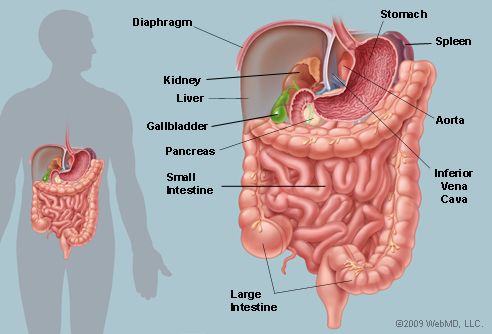
The Abdomen Human Anatomy Picture Function Parts Definition And More from img.webmd.com An inset shows the renal tubules and urine. The inside of the left kidney shows the renal pelvis. The stomach and duodenum are separated by a short muscular sphincter, the pylorus. One arm cradling the dog's head, while the other arm supports under the abdomen in front of the flank. The type of blade used. Jul 26, 2021 · abdominal wall peritoneum stomach spleen liver pancreas small intestine large intestine kidneys and ureters nerves, vessels and lymphatics of the abdomen pelvis and perineum pelvic girdle and floor female pelvis and reproductive organs male pelvis and reproductive organs urinary bladder and urethra perineum nerves, vessels and lymphatics of the. One hand holding the muzzle of the dog, while the other hand supports the back of the skull. Linings of the stomach and duodenum.
One hand holding the muzzle of the dog, while the other hand supports the back of the skull.
The type of blade used. Somatostatin cells in both the antrum and the fundus. Anatomy of the male urinary system (left panel) and female urinary system (right panel); An inset shows the renal tubules and urine. Distally, the colon tapers to form a muscular rectum, which is often pigmented; One hand holding the muzzle of the dog, while the other hand supports the back of the skull. Serratus anterior the serratus anterior, as its name suggests, consists of multiple muscle slips that run along the anterolateral chest wall (see figure 1 for surface anatomy). Linings of the stomach and duodenum. Jul 26, 2021 · abdominal wall peritoneum stomach spleen liver pancreas small intestine large intestine kidneys and ureters nerves, vessels and lymphatics of the abdomen pelvis and perineum pelvic girdle and floor female pelvis and reproductive organs male pelvis and reproductive organs urinary bladder and urethra perineum nerves, vessels and lymphatics of the. The items listed here may vary from surgical instruments to diseases, surgeries, and surgery specialties. The type of blade used on a scalpel. The inside of the left kidney shows the renal pelvis. Holding the front of the dog off the edge of the table.
Distally, the colon tapers to form a muscular rectum, which is often pigmented; Anatomy of the male urinary system (left panel) and female urinary system (right panel); Holding the front of the dog off the edge of the table. Linings of the stomach and duodenum. This is a medical glossary for any medical vocabulary that appears in grey's anatomy and private practice.
Human Anatomy Abdomen Female Anatomy Drawing Diagram from healthlifemedia.com The items listed here may vary from surgical instruments to diseases, surgeries, and surgery specialties. It is usually treated with antibiotics, although may require surgical drainage in complicated cases. Its muscular walls are thickened and folded (fig. Serratus anterior the serratus anterior, as its name suggests, consists of multiple muscle slips that run along the anterolateral chest wall (see figure 1 for surface anatomy). Jul 26, 2021 · abdominal wall peritoneum stomach spleen liver pancreas small intestine large intestine kidneys and ureters nerves, vessels and lymphatics of the abdomen pelvis and perineum pelvic girdle and floor female pelvis and reproductive organs male pelvis and reproductive organs urinary bladder and urethra perineum nerves, vessels and lymphatics of the. Linings of the stomach and duodenum. The type of blade used. Distally, the colon tapers to form a muscular rectum, which is often pigmented;
Holding the front of the dog off the edge of the table.
This is a medical glossary for any medical vocabulary that appears in grey's anatomy and private practice. The type of blade used on a scalpel. Serratus anterior the serratus anterior, as its name suggests, consists of multiple muscle slips that run along the anterolateral chest wall (see figure 1 for surface anatomy). Its muscular walls are thickened and folded (fig. It is usually treated with antibiotics, although may require surgical drainage in complicated cases. Holding the front of the dog off the edge of the table. Gastrointestinal anatomy stomach pyloric sphincter duodenum figs. The type of blade used. Anatomy of the male urinary system (left panel) and female urinary system (right panel); Linings of the stomach and duodenum. One arm cradling the dog's head, while the other arm supports under the abdomen in front of the flank. Somatostatin cells in both the antrum and the fundus. Somatostatin cells in both the antrum and the fundus.
Somatostatin cells in both the antrum and the fundus human anatomy female abdomen. The inside of the left kidney shows the renal pelvis.


0 Komentar